Fig. 6
From: Analysis of the expression of PHTF1 and related genes in acute lymphoblastic leukemia

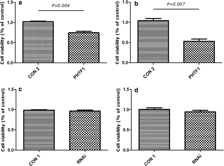
PHTF1 overexpression inhibits the proliferation of Jurkat and Molt-4 cells. a Jurkat cells infected with PHTF1 (PHTF1) or control (CON 2) lentivirus were seeded in a 96-well plate and incubated for 72 h. The CCK8 assay demonstrated that cells infected with PHTF1 have lower viability compared with control. The results shown are the mean ± SEM (n = 3). P = 0.004 for bar 1 versus bar 2. b Molt-4 cells infected with PHTF1 (PHTF1) or control (CON 2) lentivirus were seeded in a 96-well plate and incubated for 72 h. The CCK8 assay demonstrated that cells infected with PHTF1 have lower viability compared with controls. The results represent the mean ± SEM (n = 3). P = 0.007 for bar 1 versus bar 2. c Jurkat cells infected with PHTF1 shRNA (RNAi) or control (CON 1) lentivirus were seeded in a 96-well plate and incubated for 72 h. The CCK8 assay demonstrated that cells infected with PHTF1 shRNA have no significant difference compared with controls. The results represent the mean ± SEM (n = 3). NS no significance. d Molt4 cells infected with PHTF1 shRNA (RNAi) or control (CON 1) lentivirus were seeded in a 96-well plate and incubated for 72 h. The CCK8 assay demonstrated that cells infected with PHTF1-shRNA have no significant difference compared with controls. The results represent the mean ± SEM (n = 3). NS no significance